Case
A 38-year-old construction worker without significant medical history presents following witnessed syncope at her job, after standing for at least 2 hours on a particularly warm day. She reported an episode of syncope under similar circumstances 2 months prior. With each episode, she experienced “tunneling” of peripheral vision, then loss of consciousness without palpitations or incontinence. Her physical exam, vital signs (including orthostatic blood pressures), labs, and ECG were unremarkable.
Brief overview
When evaluating a patient admitted for syncope or falls, the hospitalist must address a number of questions: a) Did the patient actually have syncope?; b) What factor(s) precipitated the syncope?; c) How might similar events be prevented or mitigated in the future?; and d) Is the patient at high risk for a serious adverse outcome (for example, ventricular dysrhythmia, cardiac arrest, intracranial bleed, or death) and, therefore, in need of more immediate or intensive work-up?
The American College of Cardiology, American Heart Association, and Heart Rhythm Society guidelines define syncope as “a symptom that presents with an abrupt, transient, complete loss of consciousness, associated with inability to maintain postural tone, with rapid and spontaneous recovery” with cerebral hypoperfusion as the presumed mechanism.1 Furthermore, “there should not be clinical features of other nonsyncope causes of loss of consciousness, such as seizure, antecedent head trauma, or apparent loss of consciousness (that is, pseudosyncope).”1
A careful history revolving around the patient’s behavior prior to, during, and following the event, a thorough past medical history, and a review of current medications are essential. Potential obstacles in obtaining details of the event include lack of witnesses, patient’s inability to recall the experience, and inaccurate description of convulsive syncope as a “seizure” by bystanders.2
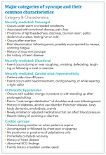
Certain characteristics may help identify types of syncope based on clinical presentation. Major categories of syncope include neurally mediated syncope (that is, vasovagal, situational, and carotid sinus hypersensitivity), orthostatic hypotension, and cardiac syncope – which may occur in the setting of acute events such as myocardial infarction, cardiac tamponade, aortic dissection, or pulmonary embolism (PE).
Overview of data
Obtaining a detailed history is crucial to understanding both the etiology of the syncopal event and determining which patients are at high risk for adverse outcomes. The etiology of syncope can be determined by history alone in 26% of patients younger than 65 years.3 Data on the prevalence of syncope by cause varies widely. As a general rule, in younger patients, especially those under 40 years of age, neurally mediated syncope is most common. As patients age, orthostatic hypotension and cardiac causes (including arrhythmias and structural diseases) occur more frequently, though neurally mediated syncope is still the most common.
Hospitalists should bear in mind that clear categorization of syncope is often challenging in the elderly. Retrograde amnesia can be seen following syncope in the aged, and even patients who can provide a history may not necessarily provide an accurate account of the event. For example, up to one half of patients who undergo tilt-table testing and have an observed episode of syncope deny that loss of consciousness ever occurred.4 Repeated falls in an elderly patient may also require an evaluation for syncope. The typical prodromal symptoms and characteristics of cardiac and neurally mediated syncope also tend to overlap in elderly patients. In a study that examined 46 variables in various age groups, only myoclonic movements during syncope and syncope during physical activity or when supine helped differentiate cardiac from neurally mediated syncope in patients over 65 years of age. Polypharmacy may also increase the susceptibility of the elderly to both orthostatic hypotension and vasovagal syncope.5 Though rare in younger patients, carotid sinus syncope should be considered in the older population, particularly under certain circumstances.
To aid the clinician in risk stratifying patients as relates to the likelihood of serious outcomes, a number of studies propose risk predictors for syncope (for example, the San Francisco Syncope Rule [SFSR], Evaluation of Guidelines in Syncope Study [EGSYS], Short-Term Prognosis of Syncope, Boston Syncope Rule, and the Risk Stratification of Syncope in the Emergency Department rule, to name a few). Unfortunately, the definition of and the timing of the adverse outcomes related to syncope often vary among studies, with reported risk factors ranging from anemia to hypotension on presentation to positive fecal occult blood testing, elevated brain natriuretic peptide, and various ECG findings. Nevertheless, several consistent predictors of serious adverse outcomes tend to emerge, such as hemodynamic instability, anemia, abnormal ECG, evidence of heart failure or structural heart disease, and acute coronary syndrome or its attendant symptoms.
Many of these predictors, however, would raise the clinical suspicion of most hospitalists for adverse outcomes in their hospitalized patients independent of the presence or absence of syncope. In fact, a meta-analysis has concluded that “None of the evaluated prediction tools (SFSR, EGSYS) performed better than clinical judgment in identifying serious outcomes during emergency department stay, and at 10 and 30 days after syncope.”6
Once the patient is hospitalized, further evaluation should be based on a careful history and physical examination. Standard evaluation also includes careful review of medications, an ECG to exclude findings suggestive of arrhythmias as well as structural or coronary artery disease, and orthostatic blood pressure measurements.1 Additional tests should be considered as deemed appropriate. For example, in patients over 40 years of age without history of carotid artery disease or stroke and in whom no carotid artery bruit is appreciated, a carotid sinus massage may be considered. The correct technique is to massage the sinus on the right then left, each for 5 seconds in both supine and standing positions with continuous heart rate and frequent blood pressure monitoring. Reproduction of syncope, especially concurrent with a cardiac pause of greater than 3 seconds and a systolic blood pressure drop of greater than 50 mmHg, is considered a positive test. Tilt-table testing should be considered in those for whom neurally mediated syncope is suspected but not confirmed, or in patients who might benefit from further elucidation of their prodromal symptoms.
If the patient’s history is concerning for arrhythmia but without supportive ECG findings, ECG monitoring should be considered. The type of monitoring will depend on the frequency of the patient’s symptoms, with consideration given to Holter monitors for more frequent events and external patch or implantable loop recorders considered in more sporadic events. An echocardiogram can be useful in those suspected of having structural heart disease. Although the overall yield of echocardiography is elucidating the cause of syncope is low,7 it may help further risk stratify those patients with suspected cardiac syncope and, in some cases, help with consideration of implantable cardioverter defibrillator placement. Cardiac stress testing may be considered for exercise-related syncope or patients suspected of having cardiac ischemia. Head imaging, EEG, and carotid ultrasounds are generally considered very low-yield in patients whose history suggests true syncope.
Of note, a study recently published in the New England Journal of Medicine suggests that the prevalence of PE in patients (median age, 80 years) presenting with a first episode of syncope was 17%, a rate that is substantially higher than historically presumed.8 Although the prevalence of PE was highest among patients presenting with syncope of unclear origin (25%), nearly 13% of patients with other explanations for syncope also had PE.


